Multiple Measurement Modes
Multiple Measurement Modes for the EnSight™
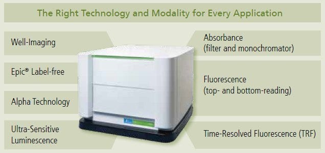
The EnSight™ Multimode Plate Reader features multiple data acquisition modalities that allow researchers to characterize several components of a single cell-signaling cascade in a single well. The fact that data are acquired and normalized on a per cell basis enable researchers to interrogate the dependence and magnitude of amplification that occurs as the signal travels downstream.
EnSight Well-Imaging Modes
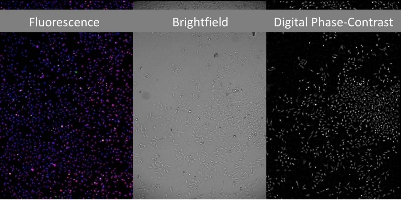
The cell-image mode of the EnSight performs at high speed with an advanced sCMOS camera that features low signal-to-background noise, laser-based autofocus and solid-state LEDs for short illumination. This modality allows users to select fluorescence, Brightfield or digital phase-content imaging modes. All these features culminate in a well-imaging module that delivers greater physiological relevance to your analysis.
Well imaging can be used to normalize data based on an individual well's population. Traditional in vivo cell based assays required researchers to use a separate assay to quantify cell number for data normalization. The well imaging technology of the EnSight automatically makes a cell number measurement and normalizes data. This powerful feature provides laboratories with a significant return on investment by reducing the number of runs.
Runs multiplex assays sequentially in a 384-well plate:
- Single color image: 4 minutes
- Two-color image: 5 minutes
- Three-color image: 6.5 minutes (approximately)
Imaging modes
- Fluorescence: enables visualization of multiple parameters per assay using up to four colors with excitation of three colors in parallel
- Brightfield: provides quick image of cells without labeling, diminishes potential labeling artifacts, cell damage, assay costs, and time
- Digital Phase-Contrast: images live cells that have not been labeled fluorescently, at greater resolution than Brightfield
Label-Free Measurement Mode
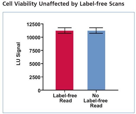
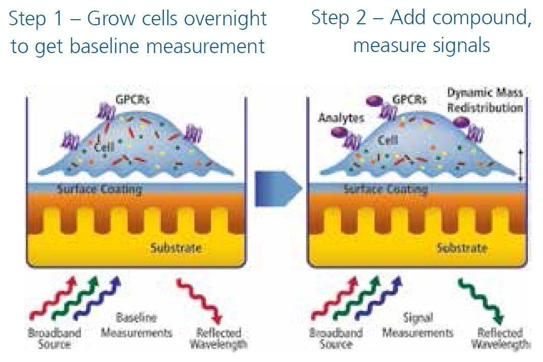
The Corning® Epic® technology for labeled and label-free imaging allows cell-based and biochemical assays to run on one platform that delivers rich, unbiased data without interference. Label-free measurement allows multiple measurements on the same plate without affecting cell viability or the need for reagents. The first image shows a cell population measured with the label-free modality and luminescence units (LU) quantified. The same cell population was then measured via the ATPLITE® luminescence based cell viability assay. The second image clearly demonstrates that the label-free measurement modality has no effect on cell viability.
Benefit: Characterizes cellular signaling mechanisms completely and with more sensitivity; use of labeled and label-free technologies provide more physiologically relevant data from target identification and screening to hit confirmation.
Applications
- Cell-based assays: Cell toxicity, Neuron biology, Phenotypic assays, Primary and stem cells, Receptor biology, Response to any stimulus, Suspension cell culture
- Biochemical assays: Aggregation, Protein- protein interactions, Small molecule binding Alpha Technology Measurement Mode
Alpha Technology Measurement Mode
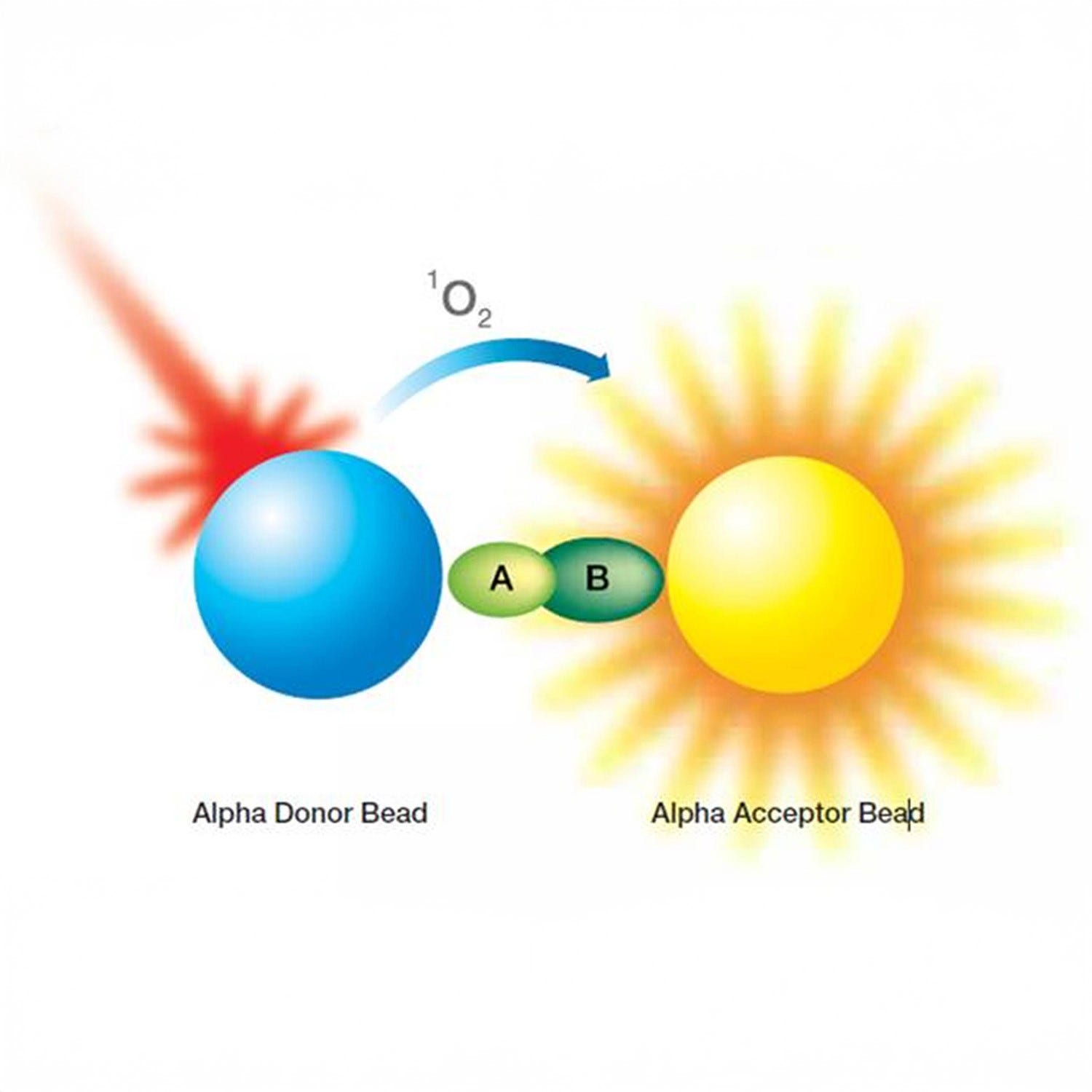
Ideal for target-based, biochemical and cell-based assays, the powerful Alpha Technology measurement modality can be used to examine virtually any line of investigation traditionally measured via an ELISA assay.
Benefits
Provided AlphaLISA® and AlphaScreen® detection produce results in half the time of standard ELISA
Applications
High-throughput analysis of bio-therapeutics, epigenetics, protein-protein interactions, G-protein coupled receptor pathways, kinase cascades and many other intracellular interactions and signaling pathways
Ultra-Sensitive Luminescence Mode
This detection mode provides robust signal strength from fewer samples by using a photomultiplier tube that boosts signal strength to a measurable level. This mode reduces the cost of assays by minimizing the amount of reagents used in each plate. Ideal for labs working with difficult-to-transfect cells, this modality measures any assay that leverages changes in luminescence.
Benefits of robust signal from fewer cells
Applications
Cellular assays, Reporter gene, Cell proliferation, Cell toxicity and viability, Flash luminescence, Primary and stem cells.
Fluorescence-Based Resonance Energy Transfer (FRET) Mode
Fluorescence-based resonance energy transfer (FRET) is a powerful tool used by researchers to interrogate the interaction of two molecules. This process links one-half of a fluorophore to each molecule of interest. If the association of the molecules is sufficiently strong, then the fluorophore will emit light at a particular wavelength when excited. This modality is extremely useful to labs studying signaling pathways, epigenetics, receptor-ligand binding, and most biochemical processes.
Benefit
Eliminates high background of other methods during detection of in vivo and in situ interactions.
Applications
GPCRs, protein-protein interactions, enzyme assays, receptor-ligand binding, kinases, endogenous receptors, cytotoxicity and cell proliferation, methylation or acetylation of peptide, histone proteins.
Time-Resolved Fluorescence and Time-Resolved FRET Mode
Highly sensitive time-resolved fluorescence measurements are used in biochemical and cell-based assays using premium and low concentration samples. TRF mode allows researchers to measure low signal intensity over time to obtain a robust signal that traditional fluorescence would fail to provide. This modality can be used to investigate a host of molecule interactions, gene expression, and other assays that would normally be measured via traditional fluorescence techniques.
Benefits
High sensitivity measurement mode ideal for low concentration samples.
Applications
GPCRs, Protein: protein interactions, Enzyme assays, Receptor-ligand binding, Kinases, Endogenous receptors, Cytotoxicity and cell proliferation, Methylation or acetylation of peptide, Histone proteins.
Fluorescence and Absorbance Mode
Equipped with a quad-monochromator component, EnSight allows you to scan on any wavelength for the best signal-to-background ratio (on fluorescence applications).
Applications
Cell counting, Colorimetric assays, GFP, ELISA, Quantification of protein and DNA/RNA, FRET, Gene expression.
