Gel Imager and Gel Documentation Features, Styles, and Applications Comparison
What is a Gel Imager? (Gel Documentation Systems)
Laboratory gel imagers, or gel documentation systems, are used by research labs to visualize and photo-document nucleic acid samples separated through gel electrophoresis, count microbial colonies, separate protein samples on western blots, and identify mixtures through thin layer chromatography (TLC).
How Does a Gel Imager or Gel Documentation System Work?
Gel imagers, or gel docs, contain an ultraviolet (UV) or visible (blue or white) transilluminator, a hood to block the samples from external light sources and a high-resolution camera for image capture.
Gel Imager Procedure
The samples, loaded onto agarose gels, petri dishes or TLC films, are placed onto the transilluminator surface. After the access doors are closed to prevent operator exposure to UV light, the lamp is powered on and the samples are photo-documented through the CCD or CMOS camera. Images are saved to the onboard software system for easy retrieval or export to Microsoft Excel or LIMS.
Where Are Gel Imagers Used?
Common in molecular biology, genomics, proteomics and microbiology labs, gel imaging systems support PCR segment identification, DNA quantification, bacterial cell culture, environmental sample testing and protein separation.
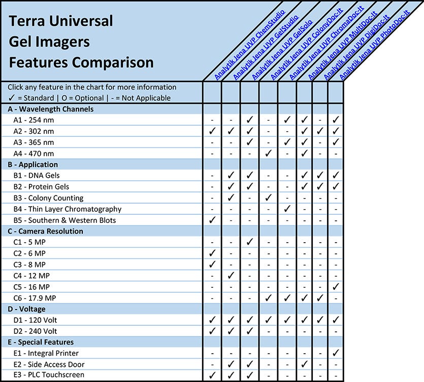
A - Gel Imager Wavelength Channels
(back to chart)
Gel imagers contain transilluminators that emit ultraviolet light or visible light for visualization of fluorescent and chemiluminescent samples or counting bacterial colonies. The UV or visible light bandwidth emitted by the transilluminator must match the absorbance spectrum of the fluorescent dye used during gel separation, blotting or thin layer chromatography.
Blue light, emitted at 470 nanometers, is optimal for colony counting or viewing samples stained with commercial dyes such as SYBR Green, SYBR Gold or SYBR Safe.
For UV visualization, 254 nanometer light is optimal for DNA cross-linking, 302 nanometer light is ideal for short exposure gels stained with Ethidium Bromide, and 365 nanometer light is best for gel band cutting.
B - Gel Imager Uses and Applications
(back to chart)
B1 - DNA Gels
Nucleic acid (DNA or RNA) samples are loaded onto an agarose or acrylamide gel and exposed to an electrical field, causing the negatively-charged samples to travel down the gel toward the positive electrode. Smaller DNA or RNA fragments travel faster than longer fragments, allowing researchers to identify fragments tagged with fluorescent dyes to excise the samples for further purification. DNA gels are commonly used for PCR analysis, cloning, and next generation sequencing.
B2 - Protein Gels
Protein samples are loaded onto a polyacrylamide gel submerged in a commercially available buffer Tris-Glycene or Tris-Acetate– Tris-Glycene or Tris-Acetate – for separation and purification. Similar to DNA gels, protein fragments of varying lengths migrate at different speeds through the gel matrix. Protein gels are used for mass spectrometry, sample denaturing, and blotting.
B3 - Colony Counting
Colony counting is the process of determining the number of microbial colony forming units (CFUs) present within the optimal growth conditions of a petri dish loaded with cell media.
What Is A Colony Forming Unit?
Each colony forming unit represents a gross estimate of the number of homogenous, viable cells growing on the plate. Colony counting is used to detect and quantify microbes in soil, water or food samples as well as identify unique cell lines for microbial cell culture research.
B4 - Thin Layer Chromatography
Thin layer chromatography (TLC) is a research technique used to separate the components of a mixture using a thin film supported by an inert backing. As the mixture moves through the column, or stationary phase, its components will migrate at different speeds based on their affinity to the column or solvent (mobile phase). Common stationary phases include silica gel, cellulose, and aluminum oxide. Common mobile phases include methanol, acetic acid, and ethyl acetate. TLC is used to quantify and qualify many substances, including lipids, carbohydrates, fatty acids, and pesticides.
B5 - Southern and Western Blot Gel
Western blotting gels, used to separate proteins for further isolation and purification, come in two primary forms: native gels and SDS-PAGE gels. Native gels, used to study enzyme or protein complexes, separate proteins based on size and ionic charge. SDS-PAGE gels, used to study antibody affinity, separate proteins based on size by denaturing the samples using the detergent sodium dodecyl sulfate (SDS).
Southern blotting gels, used to separate DNA fragments from blood or tissue samples using restriction enzyme digestion, come in two primary forms: polyacrylamide (PAGE) gels with urea and sodium dodecyl sulfate (SDS) gels with urea.
C - Gel Imager Camera Resolution
(back to chart)
Gel imagers with cameras include CCD or CMOS cameras with resolutions from 5 megapixels to 17.9 megapixels. The megapixel number represents the quantity of individual points within an image. A camera with a higher pixel count produces images with higher resolution, greater sensitivity, and better image clarity.
D - Gel Imager Voltage
(back to chart)
120-volt connections are suitable for standard laboratory power outlets in the United States.
208-volt or 240-volt connections, common in mainland Europe, require less current (amperage) and smaller conductors than equipment designed to operate at 120-volt.
E - Special Gel Imager Features
(back to chart)
E1 - Integral Printer
Although most commercially-available gel imaging systems offer optional external printers.
Analytik Jena’s UVP PhotoDoc-It systems include an integral printer for convenient printing of high-quality gel images.
E2 - Side Access Door
Analytik Jena’s GelStudio, GelSolo and MultiDoc-It imagers include side access doors for convenient loading and unloading of gels or culture plates.
E3 - PLC Touchscreen Gel Imagers
Analytik Jena’s ChemStudio, GelStudio and GelSolo imagers include integrated color touchscreens for filter and illumination control. Templates and macros, within the onboard software system, are programmable to support customized workflows.
Where Can I Buy Laboratory Gel Imagers Online?
Laboratory-Equipment.com is a specialty division of Terra Universal. For nearly 40 years, Terra Universal has served the life science, pharmaceutical, biotechnology, and medical device markets. Customers appreciate a worldwide network of reps, factory-direct support, and ready-to-ship items available from Terra's manufacturing and warehouse facilities in Fullerton, California.
Shop a wide selection of laboratory gel imagers and gel documentation systems for a wide variety of applications including general laboratory, research, PCR, DNA/RNA techniques, ELISA, protein analysis, and cell culture.
Contact a Laboratory-equipment.com specialist through web chat, email, or phone for pricing or a same-day quote.
Shop Gel Imagers by Style
Shop Gel Imagers By Compatible Stains
Terra Universal is the leading expert in the design and fabrication of ISO rated cleanrooms, furnishing and supplies.
Get a free consultation from one of our cleanroom specialists:
Call (714) 459-0731